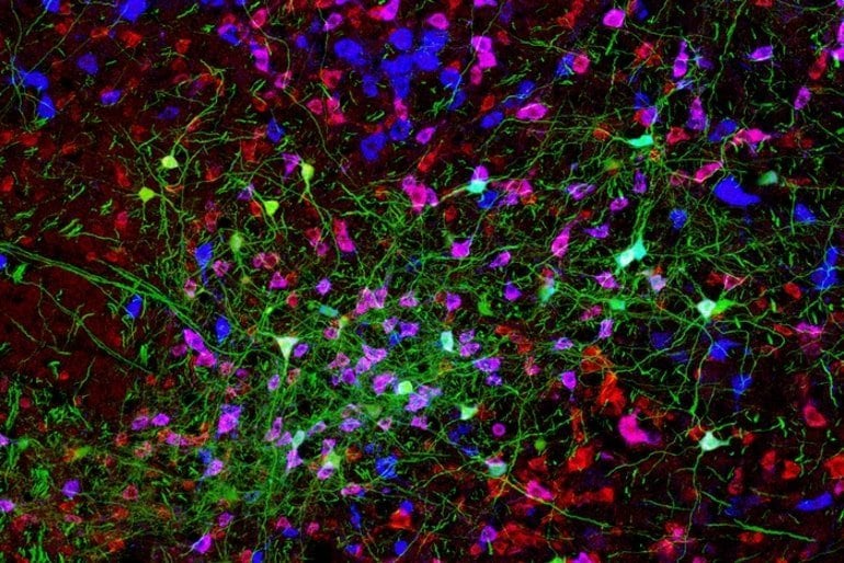
Summary: A new study in rodents reveals a beat oscillator consisting of a population of inhibitory neurons in the brain stem that fire in rhythmic bursts during beat behaviors.
Font: MIT
Many of our bodily functions, such as walking, breathing, and chewing, are controlled by brain circuits called central oscillators, which generate rhythmic firing patterns that regulate these behaviors.
MIT neuroscientists have now discovered the neural identity and mechanism underlying one of these circuits: an oscillator that controls the rhythmic back-and-forth sweep of tactile whiskers, or beats, in mice. This is the first time such an oscillator has been fully characterized in mammals.
The MIT team found that the beater oscillator consists of a population of inhibitory neurons in the brain stem that fire rhythmic bursts during the beat. As each neuron fires, it also inhibits some of the other neurons in the network, allowing the general population to generate a synchronous rhythm that retracts the whiskers from their extended positions.
“We have defined a mammalian oscillator molecularly, electrophysiologically, functionally, and mechanically,” says Fan Wang, an MIT professor of brain and cognitive sciences and a member of MIT’s McGovern Institute for Brain Research.
“It’s very exciting to see a clearly defined circuit and mechanism for how rhythm is generated in a mammal.”
Wang is the lead author of the study, which appears today in Nature. The paper’s lead authors are MIT research scientists Jun Takatoh and Vincent Prevosto.
rhythmic behavior
Most of the research that clearly identified central oscillator circuits has been done in invertebrates. For example, Eve Marder’s lab at Brandeis University found cells in the stomatogastric ganglion of lobsters and crabs that generate oscillatory activity to control the rhythmic movement of the digestive tract.
The characterization of oscillators in mammals, especially in awake behaving animals, has proven to be very challenging. The oscillator that controls gait is thought to be distributed throughout the spinal cord, making precise identification of the neurons and circuits involved difficult.
The oscillator that generates rhythmic breathing is located in a part of the brainstem called the pre-Bötzinger complex, but the exact identity of the oscillator neurons is not fully understood.
“Detailed studies have not been done in awake behaving animals, where you can record from molecularly identified oscillator cells and precisely manipulate them,” says Wang.
Whisking is a prominent rhythmic exploratory behavior in many mammals, which use their tactile whiskers to detect objects and sense textures. In mice, the whiskers extend and retract at a rate of about 12 cycles per second. Several years ago, Wang’s lab set out to identify the cells and mechanism that control this oscillation.
To find the location of the whisker oscillator, the researchers traced the motor neurons that innervate the whisker muscles. Using a modified rabies virus that infects axons, the researchers were able to tag a group of presynaptic cells for these motor neurons in a part of the brainstem called the vibrissa intermediate reticular nucleus (vIRt). This finding was consistent with previous studies showing that damage to this part of the brain eliminates the shake.
The researchers then found that about half of these vIRt neurons express a protein called parvalbumin, and that this subpopulation of cells drives the rhythmic movement of the whiskers. When these neurons are silenced, beating activity is suppressed.
The researchers then recorded the electrical activity of these parvalbumin-expressing vIRt neurons in the brainstem of awake mice, a technically challenging task, and found that these neurons peak in activity only during the period of whisker retraction. Because these neurons provide inhibitory synaptic inputs to whisker motor neurons, it follows that the rhythmic beat is generated by a constant motor neuron prolongation signal interrupted by the rhythmic retraction signal from these oscillator cells.
“That was a super satisfying and rewarding moment, to see that these cells are in fact the oscillator cells, because they fire rhythmically, they fire in the retraction phase, and they are inhibitory neurons,” says Wang.
“New Principles”
The oscillatory burst pattern of vIRt cells starts at the beginning of the beat. When the whiskers are not moving, these neurons fire continuously. When the researchers blocked the vIRt neurons from inhibiting each other, the rhythm disappeared, and instead the oscillator neurons simply increased their continuous firing rate.
This type of network, known as a recurrent inhibitory network, differs from the types of oscillators seen in lobster stomatogastric neurons, in which the neurons intrinsically generate their own rhythm.
“We have now found a mammalian network oscillator that is made up of all the inhibitory neurons,” says Wang.
The MIT scientists also collaborated with a team of theorists led by David Golomb at Ben-Gurion University, Israel, and David Kleinfeld at the University of California, San Diego. The theorists created a detailed computational model describing how the churning is controlled, which fits all the experimental data well. An article describing that model will appear in an upcoming issue of Neuron.
Wang’s lab now plans to investigate other types of oscillatory circuits in mice, including those that control chewing and licking.
“We’re really excited to find oscillators of these feeding behaviors and compare and contrast them with the whisker oscillator, because they’re all in the brainstem and we want to know if there’s any common theme or if there are lots of different ways to generate oscillators. ,” she says.
Money: The research was funded by the National Institutes of Health.
About this behavioral neuroscience research news
Author: Anne Trafton
Font: MIT
Contact: Anne Trafton-MIT
Image: The image is attributed to the researchers.
original research: Open access.
“The beater oscillator circuitby Jun Takatoh et al. Nature
Summary
The beater oscillator circuit
One-step cell surface receptors regulate cellular processes by transmitting ligand-encoded signals across the plasma membrane through changes in their extracellular and intracellular conformations. This transmembrane signaling is generally initiated by ligand binding to receptors in its monomeric form.
While downstream receptor-receptor interactions are established as key aspects of transmembrane signaling, the contribution of monomeric receptors has been difficult to isolate due to the complexity and ligand-dependence of these interactions.
By combining membrane nanodiscs produced with cell-free expression, single-molecule Förster resonance energy transfer measurements, and molecular dynamics simulations, we report that ligand binding induces intracellular conformational changes within the monomeric epidermal growth factor receptor. full-length (EGFR).
Our observations establish the existence of extracellular/intracellular conformational coupling within a single receptor molecule. We implicate a series of electrostatic interactions in conformational coupling and find that the coupling is inhibited by specific therapies and mutations that also inhibit phosphorylation in cells.
Taken together, these results introduce a simple mechanism for linking extracellular and intracellular regions via the single transmembrane helix of monomeric EGFR, and raise the possibility that intramolecular transmembrane conformational changes upon ligand binding are common to membrane proteins. in one step.

