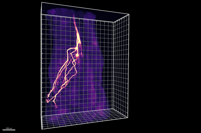
Scientists at the Scripps Research Institute have discovered sensory neurons near the spine that carry messages from fat, or adipose tissue, to the brain. This discovery debunks the prevailing notion that circulating hormones are the only messengers between fat and brain cells. The findings were published in the journal Nature (“The role of somatosensory innervation of adipose tissues“).
Nobel Laureate, Howard Hughes Medical Institute Investigator, Scripps Professor of Neuroscience and study co-senior author Ardem Patapoutian, PhD, said, “This is yet another example of how important sensory neurons are to health and disease in the body”. .”
Li Ye, PhD, associate professor of neuroscience, chair of chemistry and chemical biology at Scripps and lead author of the study, said, “The discovery of these neurons suggests for the first time that your brain is actively examining your fat, rather than simply passively receiving messages about it. The implications of this finding are profound.”

In addition to harboring long-term energy reserves and releasing it when needed, adipose tissue regulates hormones and signaling molecules that update the brain on satiety and metabolism. Disruption of these crucial functions of adipose tissue causes or contributes to several metabolic diseases, including diabetes, fatty liver diseaseatherosclerosis and obesity.
Scientists previously suspected that the nerves extending into adipose tissues were part of the sympathetic autonomic nervous system that activates fat-burning metabolic pathways during physical activity or stress. The lack of adequate research techniques to study the innervation of adipose tissue has so far prevented researchers from identifying the nature and function of neurons in fat cells. Conventional methods used to visualize or stimulate neurons in the brain or near the body surface do not work on fat cells.
A tissue-cleansing technique developed by Ye and colleagues called Hybrid, uses solvents to dissolve opaque biomolecules like fat to make tissues transparent. In the current study, Ye’s team applied this protocol to track neurons that infiltrate deep into adipose tissue. They found that almost 50% was transmitted not to the sympathetic ganglia that flank the spinal cord, but to the dorsal root ganglia (DRG), a group of cell bodies of sensory neurons. The technique allowed the team to visualize the entire axonal projection of DRG from the cell body (soma) to fat cells under the skin (subcutaneous adipocytes) and to establish its anatomical origins.
Next, to assess the function of these DRG neurons in adipose tissue, Ye’s team used a genetic technique previously developed by the team called ROOT (retrograde vector optimized for organ tracking). This allowed them to measure the consequences of removing selected small sets of sensory neurons within adipose tissue.
Study first author Yu Wang, a graduate student in Ye and Patapoutian’s labs, said: “This research was really made possible by the way these new methods came together. When we started this project, there were no tools to answer these questions.”
The researchers found that experimental removal of adipose tissue sensory inputs to the brain in mice triggers the generation of fat (lipogenesis) and increases the conversion of white fat to brown fat, resulting in enlarged pockets of fat under the skin. and elevated body temperatures under normal environmental conditions. temperatures High levels of brown fat are known to break down other fat and sugar molecules to produce heat.
These results suggest that sensory and sympathetic innervation in adipose tissue may play opposing roles: while sympathetic neurons trigger fat burning, sensory neurons trigger fat production. Ye said: “There is no single instruction that the brain sends adipose tissue. It’s more nuanced than that. These two types of neurons act as an accelerator and a brake to burn fat.
Among the questions that remain unanswered is the nature of the messages to and from adipose tissues. Ye’s team is currently taking a closer look at sensory signals in fat cells and looking for similar sensory neurons in visceral organs.
